What are X-rays, how were they discovered, and how much impact do they have on humans?
X-rays are a type of light, so they are also called X-rays.

X-rays are a type of light, so they are also called X-rays.
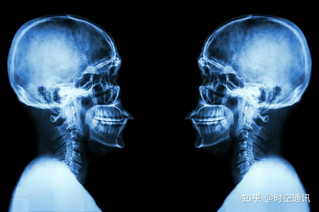
When people go to the hospital for fluoroscopy and CT scans, they use X-rays, also called X-rays. X-rays are actually a kind of light, but this kind of light is different from the light we usually know. It can pass through certain objects behind the human body, but the eyes cannot see it.

Light is actually the transfer of energy, and its essence is a photon flow in a specific frequency band. The light source emits light because the electrons in the light source gain extra energy and release energy in the form of waves during the transition process.
This is true for sunlight, electric light, and firelight. Therefore, light is essentially an electromagnetic wave that relies on photons to transmit energy information. However, there are visible and invisible light. In the long-term evolution of the human eye, it is only sensitive to the frequency band of about 380~780nm, so the electromagnetic waves in this specific frequency band are called visible light.
In addition to visible light, there are many types of light that are invisible to the human body, such as radio waves, infrared rays, ultraviolet rays, X-rays, and gamma rays, which are invisible light. These lights are all in a certain band and frequency in the electromagnetic spectrum.
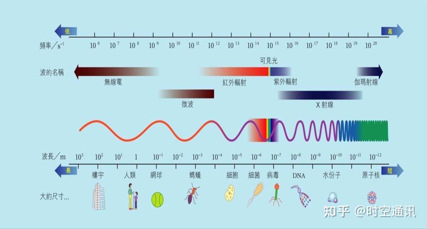
These invisible lights have low and high energies, and visible light is in the middle band. Light with lower energy than visible light includes radio waves (including long waves, medium waves, short waves, microwaves) and infrared rays; light with higher energy than visible light includes ultraviolet rays, X-rays, and gamma rays.
X-rays are electromagnetic waves second only to gamma rays, with wavelengths between 10 nanometers and 0.01 nanometers, frequencies between 3^16 and 3^20 Hz, and energies between 124eV and 1.24MeV. This is the energy of each photon, and it is a high-energy ray, so it has strong penetrating power. When X-rays irradiate the human body, part of it is absorbed by human body matter, and most of it passes through the gaps between atoms.
The higher the frequency and the shorter the wavelength, the greater the X-ray energy and the stronger its penetrating ability. In the process of penetrating an object, the absorption is different according to the density and thickness of the object, so the X-rays that pass through are strong or weak, thus showing the structure of the object on the photographic film.
X-rays were discovered accidentally in 1895
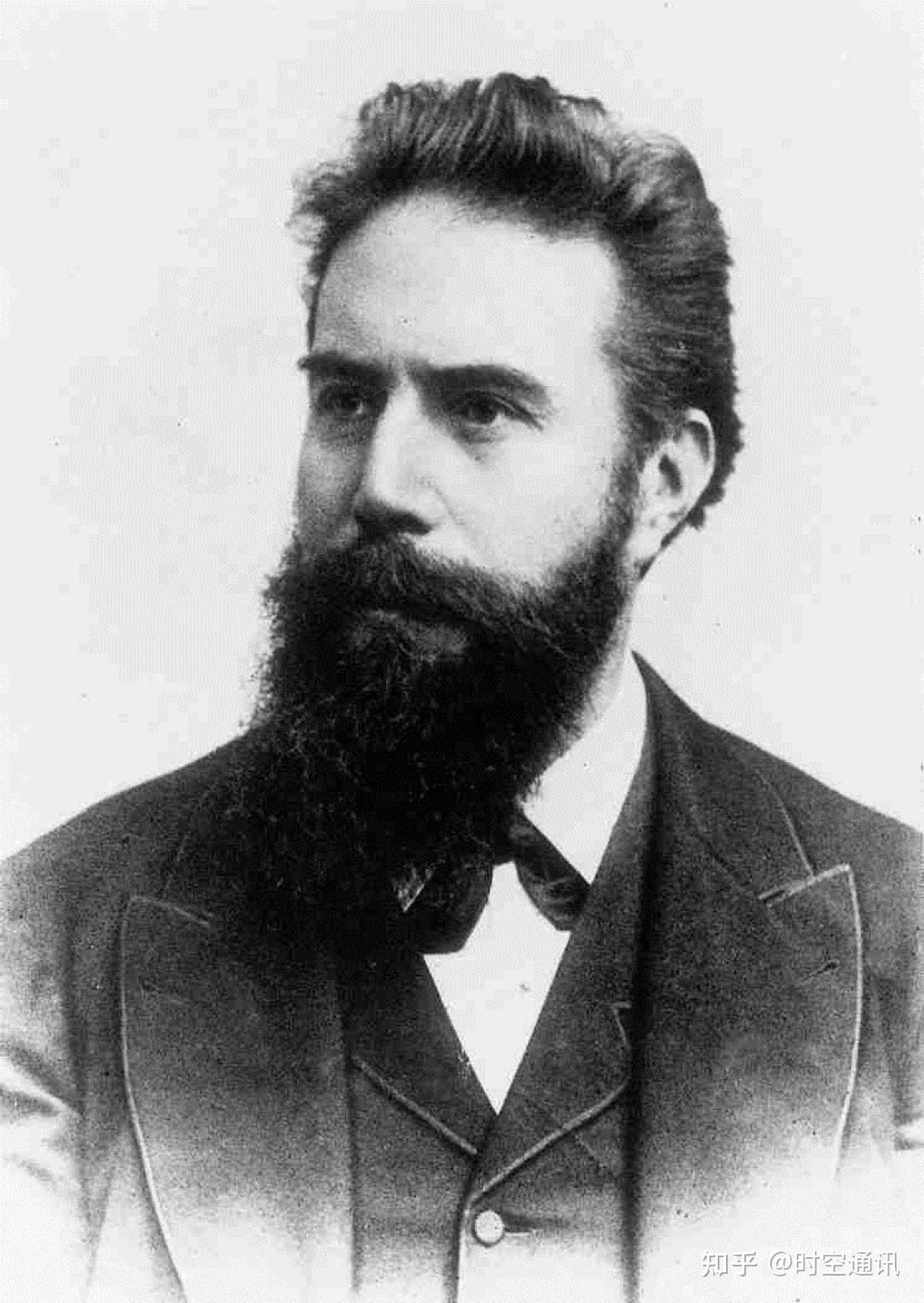
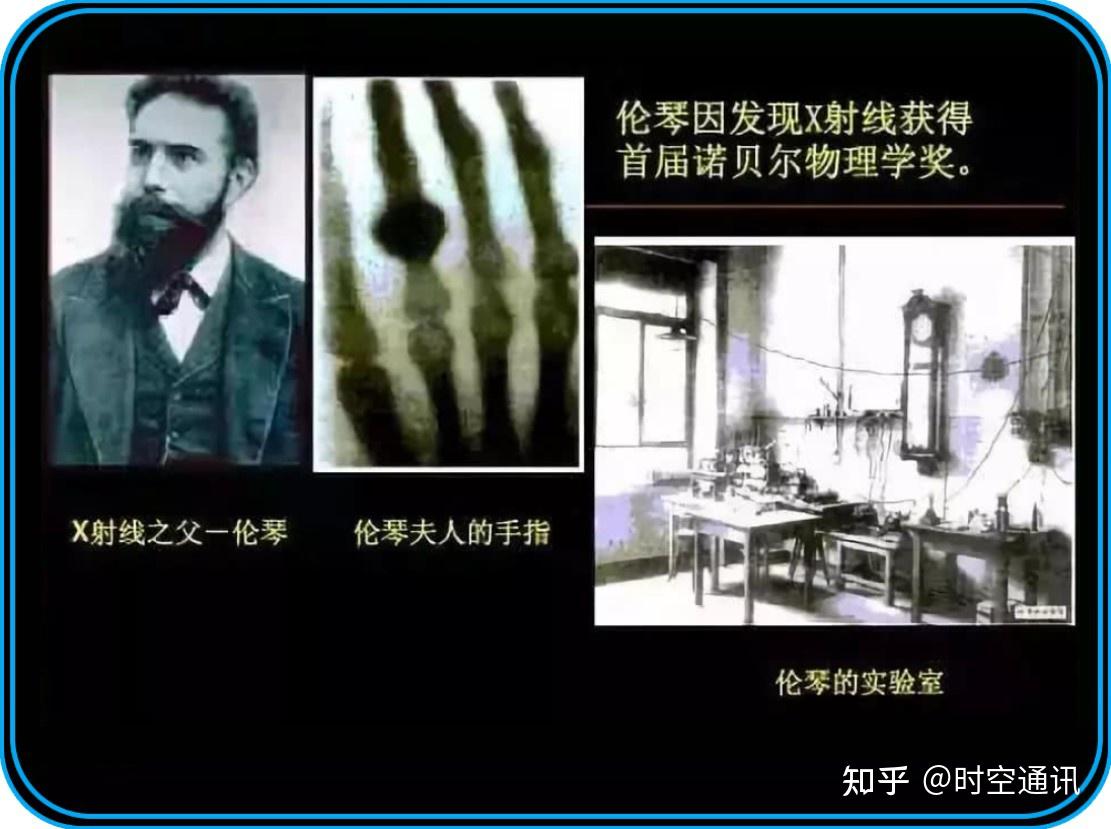
When talking about the discovery of X-rays, we have to mention a great scientist, Wilhelm Conrad Röntgen . Röntgen was born in 1845 in a relatively well-off family of small business owners in the city of Rhineland-Palatinate, Germany. He received a good education since childhood and once studied under the famous thermodynamics scientist Clausius . In 1868, he was hired as a professor at the University of Würzburg .
Later, Roentgen went to many universities to conduct physics research. In 1888, he returned to the University of Würzburg. Soon after, he succeeded Konte as director of the Institute of Physics. In 1894, he was elected as the president of the university.
At the end of the 19th century, Europe was a time of surging science. Many European physicists were obsessed with studying vacuum discharge phenomena and cathode rays, and Roentgen was also deeply involved in it. However, Roentgen’s research was more sophisticated. In order to prevent the influence of external light on the discharge tube and prevent the visible light in the tube from leaking out of the tube, he darkened the entire room and made a black cardboard cover to tightly seal the discharge tube.

But when he connected the high voltage to test the cathode ray, the black cardboard cover did not emit a single light, and he was satisfied with his masterpiece. But at this moment, he suddenly found a little flash 1 meter away from the discharge tube, and the flash disappeared after cutting off the power. It was a fluorescent screen coated with barium cyanide platinum, and it would emit a faint light green flash every time the power was turned on!
He was surprised, because he knew it was definitely not cathode rays, because he and many other scientists had confirmed through experiments that cathode rays could only move a few centimeters in the air. He tried to move the fluorescent screen farther away, until it was 2 meters away, and it still emitted flashes.
At this time, Roentgen was ecstatic. This was probably something that had never been discovered before, an unknown ray! But he couldn’t rule out whether this was an illusion or phantom of his eyes in the dark. So in the next few weeks, in order to confirm the existence of this ray, he locked himself in the laboratory and studied it silently.
Because science is a very rigorous thing, and making claims without verification goes against the scientific spirit, he didn’t tell anyone.
He placed a small bed in the laboratory and asked someone to bring food to him. In this way, he ate and lived in the laboratory, so that the experimental equipment could not be interrupted and the test could be continuous. In order to eliminate the illusion of vision, he used a photographic plate to record these strange lights. Later, he used paper, books and wooden boards to separate the photographic plates. These rays passed through unyieldingly, as if they were transparent to it.
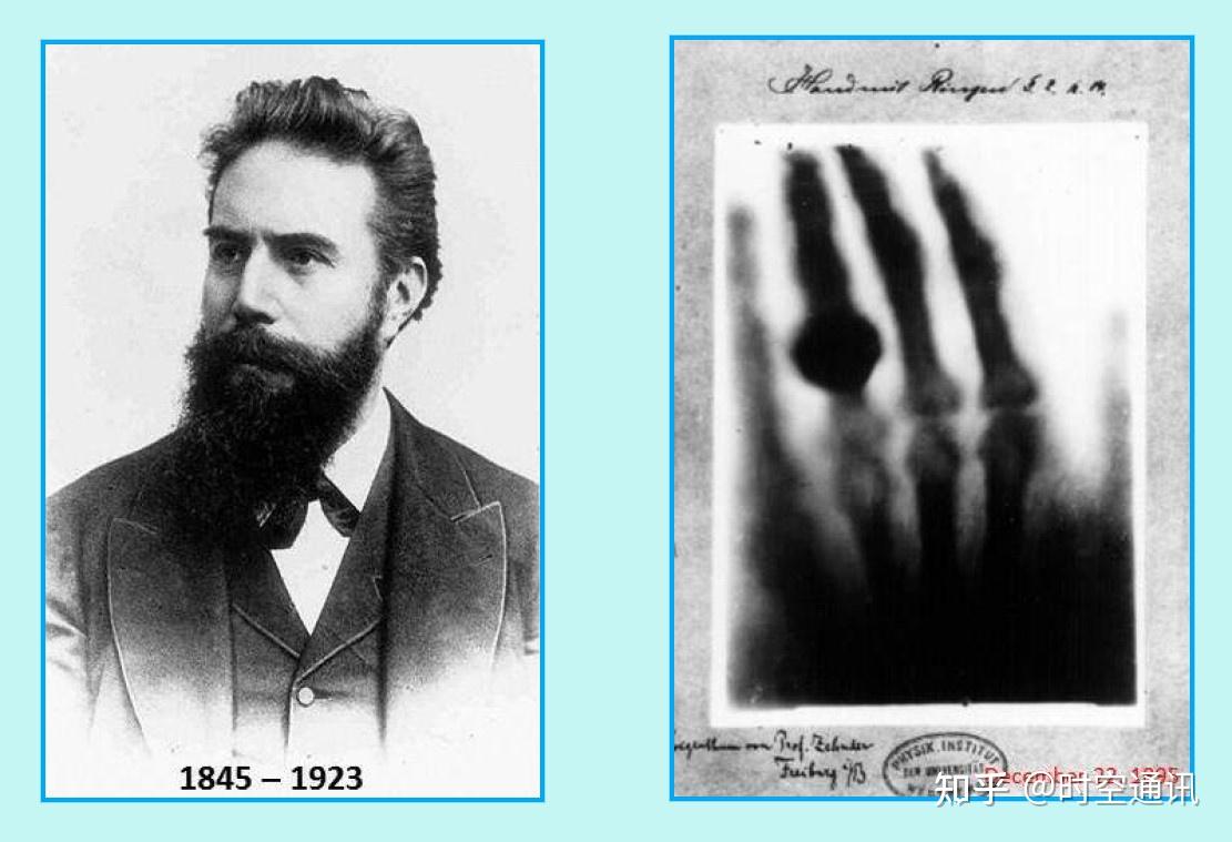
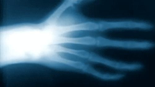
Seven weeks had passed when he felt that the new light was certain. On the night of December 22, 1895, he persuaded his wife to be the subject of the experiment. When his wife stretched herself with a ring on her body toward the fluorescent screen, a shocking and strange phenomenon appeared. Mrs. Roentgen saw a bony hand. Looking more closely, it was not a hand, but a series of joints.
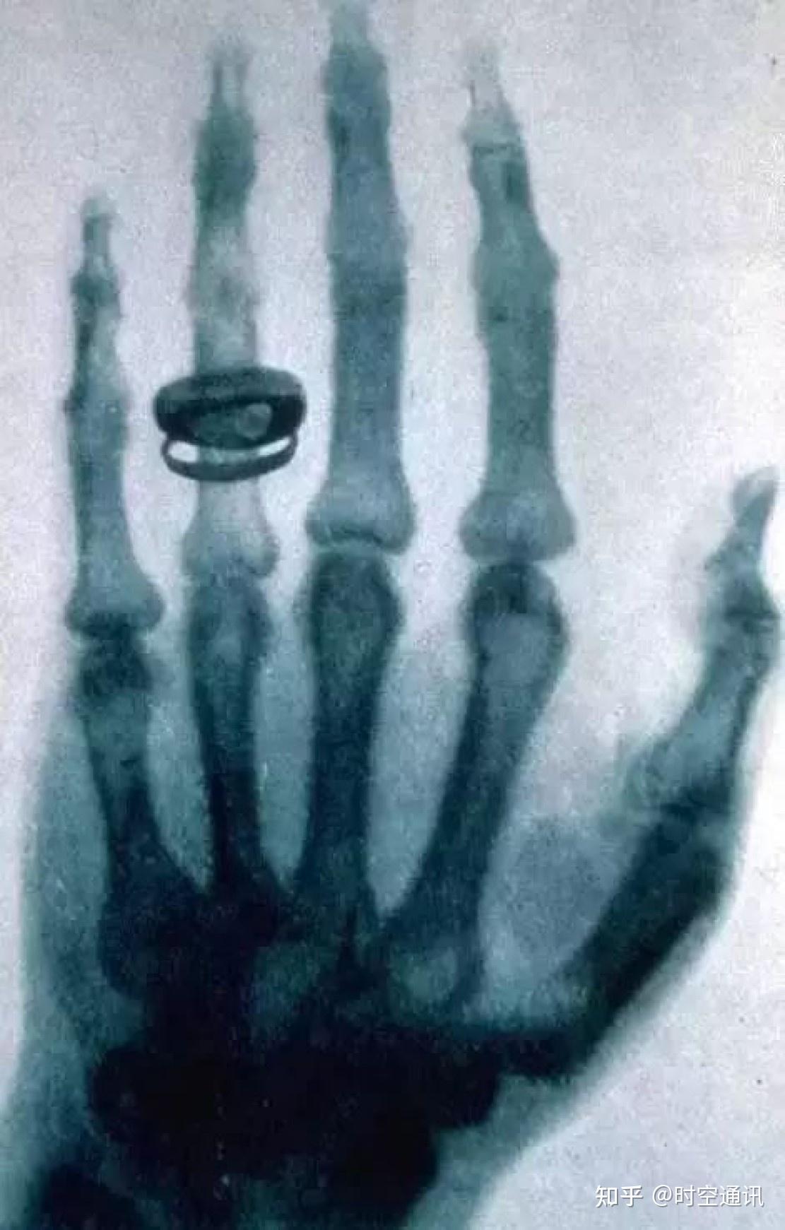
Mrs. Roentgen couldn’t believe it. Was this her hand? But on the shadow’s ring finger, there was clearly a ring, and the position of her ring was exactly the same as hers! Mrs. Roentgen was frightened by her own hand. Who else in that era had seen such an image? Only the bones of the dead could be like this.
This was the first X-ray photo of a human body! Roentgen was so excited that he hugged his wife. It was finally confirmed that this was an unprecedented type of light that could penetrate the flesh.
Roentgen’s discovery shocked the world
On December 28, 1895, Roentgen submitted his paper “A New Ray, Preliminary Report” to the Wurzburg Physical and Medical Society, in which he named the ray with the symbol “X”. He said that when I discovered this phenomenon, it was so strange and amazing that I exhausted myself, doing the same experiment again and again to exclude illusions or phantoms. For several weeks, I didn’t want anything else to interfere with my experiment.
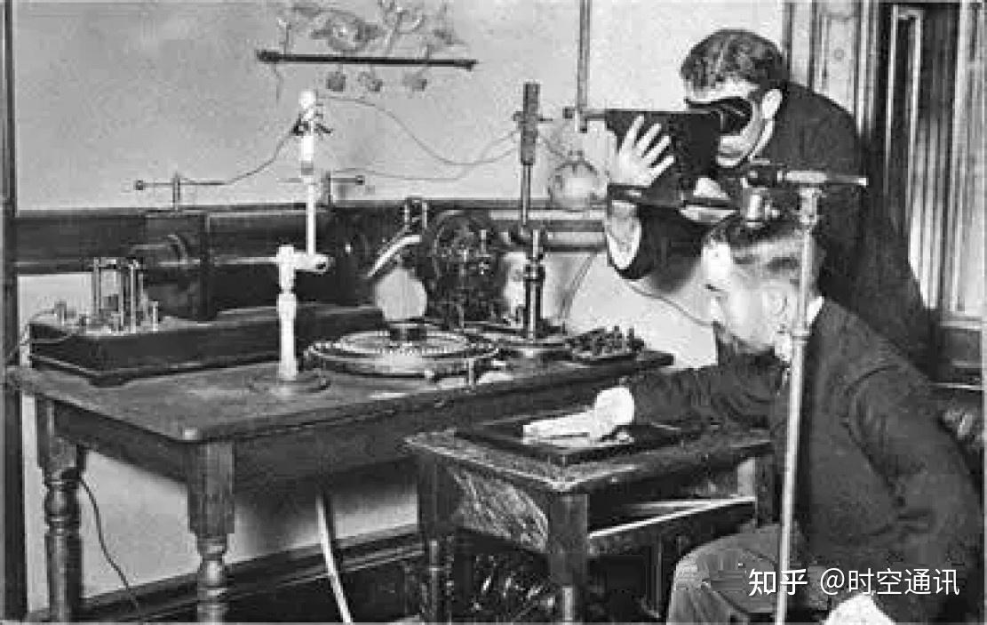
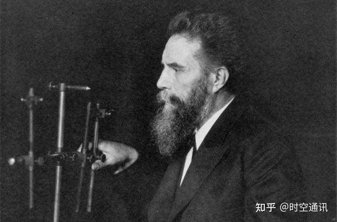
Roentgen was a low-key person, but on January 23, 1896, he held the only report conference in his life at his institute and announced his discovery. At the report conference, Roentgen asked the famous anatomist of the University of Würzburg, Crickel , to extend his hand so that he could take an X-ray on the spot. Crickel readily agreed. When the dry plate was developed and the beautiful hand bones of an 80-year-old man appeared, the audience burst into applause and cheers.
Crickel immediately proposed excitedly to name this kind of ray “Roentgen ray”. Later, people called the radiation dose unit of X-ray and gamma ray “Roentgen”. Since then, Roentgen’s discovery has spread all over the world, causing a huge sensation that has never been seen in the world.
All research institutions rushed to copy Roentgen’s experimental equipment and repeat his experiments. Of course, real scientific achievements can withstand any duplication. X-rays became a fashion all over the world.

Some people took photos with X-rays, and it was fashionable to take photos of skeletons. Even shoe merchants used it to promote sales, using X-rays to try on shoes. At that time, people did not know the harm of X-rays at all, and thought it was a wonderful feeling brought to humans by God.
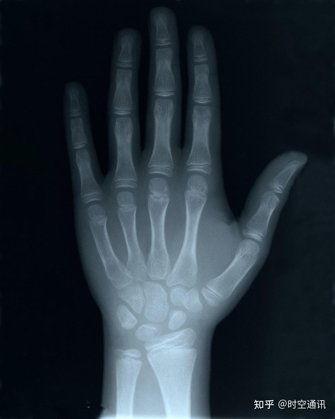
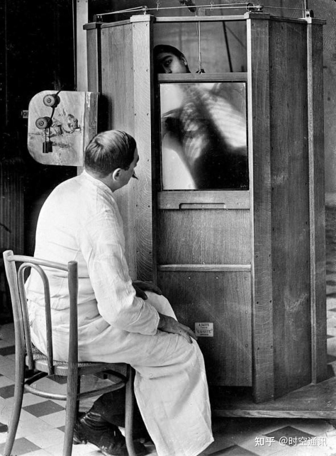
The earliest application of X-rays was in medicine. This is the perspective technology. Through X-rays, people can see lesions that should only be seen through dissection, which has better relieved the pain of countless people. At that time, Thomas Henry, a famous British surgeon , called X-rays “the greatest milestone in the history of diagnosis.”
Roentgen’s keen vision and rigorous scientific attitude made it possible to discover X-rays, which has profoundly changed human life. Due to Roentgen’s great contribution, he won the first Nobel Prize in Physics in human scientific history in 1901.
But Roentgen kept a low profile throughout his life. He did not apply for patents for his inventions, nor did he refuse the noble title bestowed on him by German Emperor Wilhelm II. He continued to work silently on his science as an ordinary person, and achieved achievements in many fields such as optoelectronics, heat, and electromagnetism. He won more than 150 awards in his lifetime, but they were all overshadowed by the discovery of X-rays.
On February 10, 1923, Roentgen died in Munich, but his spirit lives on.
Science is a neutral double-edged sword
The discovery of X-rays has given the world a great stimulus. People are immersed in the ecstasy of discovery and widely use X-rays in various occasions of social life. At first, people did not realize the harm of this kind of rays. With the abuse of X-rays, its harm gradually appeared.
At the end of January 1896, American Gruber was injured by radiation while making X-ray tubes and conducting X-ray experiments, and eventually his fingers and parts of his palms were amputated; in March 1896, American Edison felt eye pain while making an X-ray fluoroscopy device, and then developed conjunctivitis; in April 1896, American Daniel discovered that X-rays could damage hair and cause it to fall off when he used X-rays to irradiate the location of foreign bodies in the head; in July 1896, German Matthews described the hair loss and dermatitis caused by X-ray fluoroscopy.
But people still did not realize the seriousness of the situation. Some people even used large doses of X-rays to remove body hair for women. Later, these women developed wrinkles, spots, infections, ulcers and skin cancer to varying degrees. From 1930 to 1960, the medical community regarded X-ray fluoroscopy as the most fashionable diagnostic and treatment method. Some patients developed malignant tumors such as leukemia, bone tumors, and liver cancer due to high-dose cumulative radiation.
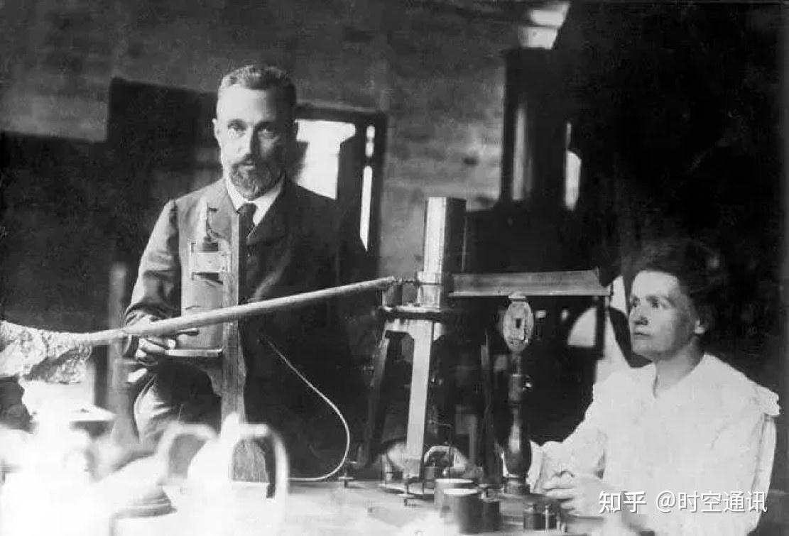
In fact, Roentgen, the discoverer of X-rays, was also a victim of X-rays. He died of multiple visceral cancers due to years of exposure to X-rays. Marie Curie , a Nobel Prize winner who also worked with radioactive elements throughout her life , also died of cancer due to radiation (the picture above shows the Curies).
It was not until the 1960s, as more and more X-ray injury incidents occurred, that people truly realized the dangers of this type of radiation and began to pay attention to protection and use it in safe doses.
Radiation dose unit
Nowadays, X-rays are widely used in various aspects of society, especially in medicine and industrial flaw detection. So how much X-ray exposure dose is safe for humans?
To clarify this question, we must first understand the radiation dose units. The main radiation measurement units are: R (roentgen), rem (rem), Sv (sievert), mSv (millisievert), μSv (microsievert), etc., and the commonly used ones are Sv, mSv, and μSv.
Sv is a relatively large unit. 1Sv exposure means that the human tissue absorbs 1Gy (Gray) of radiation, and also obtains 1J/kg (Joule/kilogram) of radiation. This radiation dose is very large, equivalent to the radiation dose received by the survivors of the Hiroshima atomic bomb explosion.

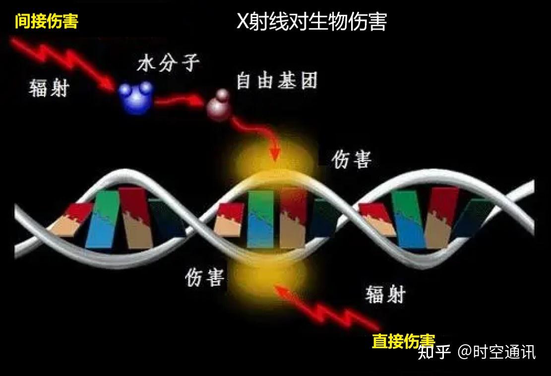
The radiation dose unit does not only refer to X-ray radiation, but also includes other radiation, such as gamma rays, etc. These high-energy rays have high energy and can penetrate biological organisms, break DNA molecular bonds, damage or even kill cells, cause damage to the body, and large doses of radiation can even cause immediate death.
The relationship between these units of measurement is: 0.01Sv (Sievert) = 1R (roentgen) = 1rem (rem) = 10mSv (millisievert) = 10,000μSv (microsievert).
Safe and lethal doses of radiation
In fact, radiation is everywhere around us. According to the United Nations Scientific Committee on the Effects of Atomic Radiation, based on the natural radiation exposure in various parts of the world, the average natural radiation dose received by each adult is about 2.4mSv per year. This 2.4mSv includes 0.4mSv of cosmic rays, 0.5mSv of ground gamma rays (external radiation), 1.2mSv of inhalation (mainly radon), and 0.3mSv of ingestion (internal radiation).

Research shows that a single exposure of 4.5 Sv is a half-lethal dose, which means a mortality rate of about 50%, which is equivalent to 300 to 400 consecutive CT scans; a single exposure of 6 Sv has a mortality rate of almost 100%. After long-term observation and research, the annual exposure of the human body to X-rays does not exceed 50 mSv, which is within the safe tolerance range.
When people receive X-ray or CT scans in the hospital, they are also exposed to X-rays. The radiation dose each time varies depending on the part of the body, and ranges from 0.01 to 15 mSv. Even with all kinds of protection, astronauts in space are exposed to 100 to 200 times more radiation than on the ground. People who smoke a pack of cigarettes a day are exposed to about 10 to 50 mSv of radiation per year, which is equivalent to 50 chest X-rays, or the amount of radiation a non-smoker would receive in 5 to 20 years.

In life, we may encounter many radiation environments. We must take precautions to make good use of radiation, a scientific double-edged sword, and serve the progress of human civilization. Thank you for reading and welcome to discuss.
